|
|
|
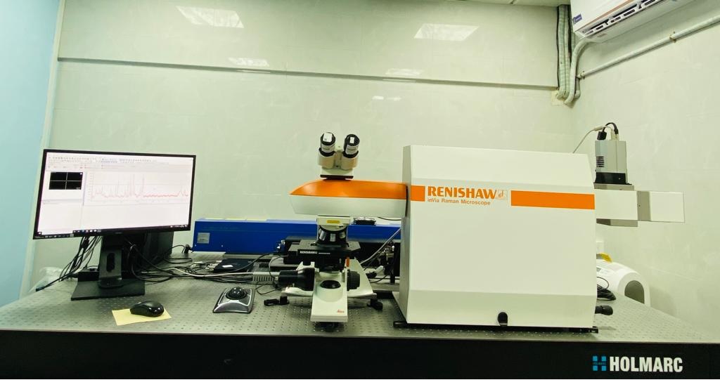
Laser Raman Imaging System (LRIS)
|
 Register your request here (Internal Users) and (External Users) and submit the hardcopy of filled Material Safety Data sheet along with sample. Register your request here (Internal Users) and (External Users) and submit the hardcopy of filled Material Safety Data sheet along with sample.
| Make |
: Renishaw, UK |
| Model |
: Invia Reflex
|
 |
| Specification |
:
 Spectrograph equipped with a research-grade microscope capable of producing Raman Spectrograph equipped with a research-grade microscope capable of producing Raman
(wavenumber transfer 50 to 4000 cm-1) and PL (330 nm to 1.6 microns).
Spectral Range of spectrometer: 200 nm – 1600 nm
Excitation sources-
| Laser available | HeCd 325 nm | Diode 532 nm | HeNe 633 nm | Diode 830 nm |
| Power | >25 mW | >50 mW | >17 mW | >200 mW |
| Spectral resolution (FWHM) | 2 cm-1 with 2400 gr/mm | 0.5 cm-1 with 2400 gr/mm | 1 cm-1 with 1800 gr/mm | 0.75 cm-1 with 1200 gr/mm |
| PL | 330 nm to 1600 nm |
|
Microscope Objective- Normal WD-5X,20X,100X
Long WD-50X
NUV-15X & 40X
Imaging/Mapping-
XYZ motorized and computer-controlled mapping stage with a minimum travel distance of 110x75x25 mm with a resolution of
50x50x10 nm. Scanning step size for 2D mapping is better than 50 nm.
Temperature- 10 to 300 K (CCR), Room temperature to 1500 K (Furnace)
Libraries- Inorganic and minerals, polymeric materials, biochemical.
Other capabilities- Low wavenumber measurements (down to 15cm-1 with 532 nm laser),
polarization study with 532 nm laser, remote probe fibre coupling with 532 nm laser.
|
|
The Raman effect is based on scattering of light, which includes both elastic (Rayleigh) scattering at the same wavelength as the incident light, and inelastic (Raman) scattering at different wavelengths, due to molecular vibrations. Raman scattering is about a million times less intense than Rayleigh scattering. Therefore, to obtain Raman spectra, it is necessary to prevent Rayleigh scattering from overpowering the weaker Raman scattering.
Raman spectra are measured by exciting a sample using a high-intensity laser beam, with the resulting scattered light being passed through a spectrometer. The Raman shift is the energy difference between the incident light and the scattered light. In the resulting spectrum, the vertical axis is the intensity of the scattered light and the horizontal axis is the wavenumber of the Raman shift (cm-1).
The Raman shift is associated with two different energy bands. The shift at wavelengths higher than that of the incident light is termed Stokes scattering. The shift at wavelengths lower than that of the incident light is termed anti-Stokes scattering. Stokes scattering is observed in the lower wavenumber (longer wavelength) region and anti-Stokes scattering in the higher wavenumber (shorter wavelength) region. Typically, higher-intensity Stokes scattering peaks are used for analysis.
|
 Pharmaceuticals and Cosmetics Pharmaceuticals and Cosmetics
Geology and Mineralogy
Carbon Materials
Semiconductors
Life Sciences
Polymer science
|
LRS Lab, 1st Floor,
CRNTS/SAIF, IIT Bombay
|
|
|
|
Prof. Anil Kottantharayil
Contact : 022-25767438
Email Id : anilkg@ee.iitb.ac.in
|
Dr. Mayuri Gandhi
CRNTS/SAIF IIT Bombay
Contact : 022-2159 6899
Email Id : lris@iitb.ac.in,pallaviag@iitb.ac.in,mngandhi@iitb.ac.in
|
|
Laser Raman Imaging System Charges (18% GST will be applicable on below specified charges for external users)
|
| | Industry | University | National Lab/R&D's | IIT Bombay Users | |
| Raman Analysis | 1800/- | 600/- | 1200/- | 300/- | Per Sample |
| T-dependent studies (cryostat 10 to 300 K)† | 6000/- | 1200/- | 3000/- | 600/- | Per Slot |
| T-dependent studies (furnace 300 to 1500 K) | 4500/- | 800/- | 2250/- | 400/- | Per Slot |
| Specialized measurements (measurements up to 15 cm-1, polarization measurements) | 4000/- | 800/- | 1600/- | 400/- | Per Slot |
| Raman Analysis (Imaging/mapping) | 5000/- | 1000/- | 2000/- | 500/- | Per Slot
(1 hr 30 min) |
|
† subject to revision based on cost incurred
|
|